Blog post #2 by Ellie Kimura
I have been working in my lab for about three weeks and it has been an amazing experience full of learning new technical skills in the lab and making new connections with people from all around the world.
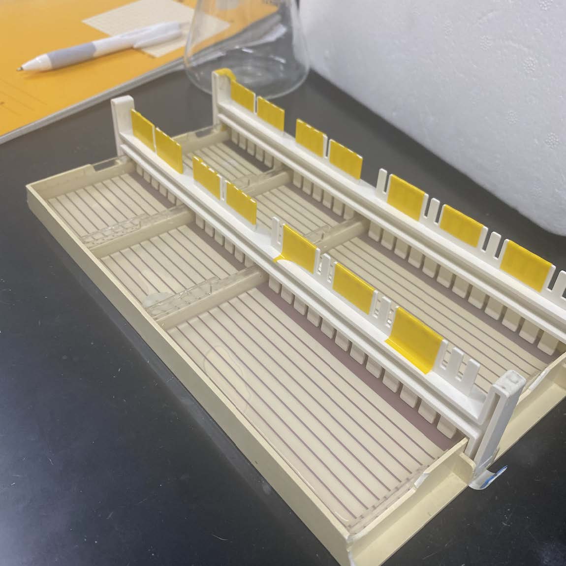
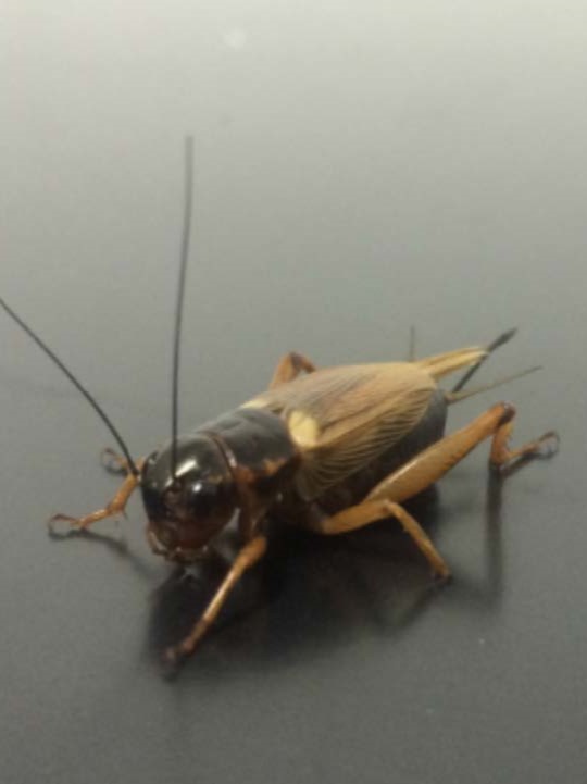
For the first two weeks in my lab, I was mostly spending my time practicing my cricket dissection technique in order to become competent enough to cleanly dissect all the major organs from the cricket. This was definitely my biggest challenge, as it took hours of looking through a microscope and performing many failed dissections. I found the brain to be particularly difficult to dissect, as it is extremely small and fragile. I would say that my biggest achievement was when I successfully dissected the brain, subesophageal ganglion, fat body, midgut, malpighian tubules, and accessory gland from a single cricket earlier this week. This was a significant achievement for me because the ability to dissect these organs was essential in order for me to proceed to the next step of my experiment, which is RNA extraction, followed by a Reverse Transcription of RNA to cDNA. The next step of my experiment is to conduct a RT-PCR using a specific primer for an insulin-like peptide in order to gain a better understanding of the specific organs that contain insulin-like peptides and how this may affect feeding behavior and endocrine functions of the cricket.